Carbon Design Innovations has announced that it has grant in the amount of $390,000 from the National Institutes of Health (NIH) Small Business Innovation Research (SBIR) Program. The grant will fund the development and commercialization of Carbon Nanotube (CNT) Atomic Force Microscope (AFM) probes for bioimaging and investigations in cellular biology. Carbon Design Innovations will collaborate with the University of California at Davis, US on the development of the probes.
www.carbondesigninnovations.com
Grant from NIH to Develop AFM Probes
August 19, 2009Canada Gains New Centre for Nanotechnology
Juli 20, 2009Alberta, Canada will soon be home to a new research and product development centre for nanotechnology called Hitachi Electron Microscopy Products Development Centre (HEMiC) at the National Institute for Nanotechnology (NINT) in Edmonton. The centre will house three new electron microscopes valued at $7 million. The $14 million project is supported by the Western Economic Partnership Agreement between the Governments of Canada and Alberta and to contributions from Hitachi High-Technologies. The HEMiC is made possible by a wider collaboration of the Alberta Ingenuity Fund’s nanoWorks program, the National Institute for Nanotechnology of the National Research Council, the University of Alberta and Hitachi High Technologies Canada Inc. One of the centre’s first projects will evaluate and test the world’s sharpest electron emitter, developed by the Molecular Scale Devices group at NINT for use as an electron source in electron microscopes.
www.nrc-cnrc.gc.ca
State-of-the-Art Geosciences Laboratory Opened
Juni 29, 2009An advanced science laboratory has officially been launched on June 26, 2009 at the University of Monash, Victoria, Australia by the Minister for Innovation, Industry, Science and Research Kim Carr. The $1 million Earth Sciences teaching laboratory will provide students with the latest in high-tech learning, giving them access to next-generation computer modelling and microscope technology. Head of Geosciences School Professor Ray Cas said the laboratory had the capacity to teach at a microscopic scale via the linking of microscopes with the smart screens. „The laboratory is the most advanced facility of its kind in Australia and the technology it employs is at the cutting-edge internationally,“ Professor Cas said.
www.monash.edu.au
From Tweezers and Microscopes
Juni 19, 2009The registration for the eighth annual symposium on the applications of scanning probe microscopy (SPM) actually opened. Along with the meeting goes the second annual symposium on optical tweezers. The symposia will be held on the October 14-15, 2009 in Berlin and will focus on applications developments in life sciences. These meetings have become highly regarded on the international SPM meetings calendar. JPK again expects more than 100 scientists from around the world to come to Berlin to discuss their results and share scientific knowledge. To learn more or to attend this meeting, visit:
www.nanobioviews.net
www.jpk.com

Eighth annual symposium on the applications of scanning probe microscopy (SPM)
$2 Million Grant for Live Microscopy
Mai 13, 2009A proposal by a team of UC Davis (University of California, US) scientists to develop the first electron microscope capable of filming live biological processes has been awarded a $2 million grant from the National Institutes of Health. The team’s plan is to extend the capabilities of a powerful new imaging tool called the dynamic transmission electron microscope or DTEM. These instruments can snap 10 to 100 images per millionth of a second, while capturing details as small as 10 nanometers. If they can be adapted to living, moving systems, DTEMs could achieve resolutions 100 times greater than currently attainable for live processes, enabling scientists to observe and record biological processes at the molecular level. Currently, there are only three DTEMs in use worldwide, none of which are designed for observing living systems. Rather, they are utilized to document such processes as inorganic chemical reactions and the dynamics of materials as they change from one state – solid, liquid or gas – to another.
www.ucdavis.edu
Live Cell Imaging at Double the Resolution
Mai 6, 2009A team of researchers of the University of Georgia (UGA) and the University of California, San Francisco, US has developed a microscope that is capable of live imaging at double the resolution of fluorescence microscopy by using structured illumination. The research was published in Nature Methods on April 26, 2009. “What we’ve done is develop a much faster system that allows you to look at live cells expressing the green fluorescent protein (GFP), which is a very powerful tool for labeling inside the cell,” explained UGA engineer Peter Kner.
www.engineering.uga.edu
The Smallest Periscope
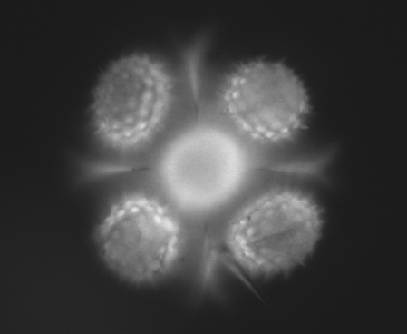
März 11, 2009A team of Vanderbilt scientists have invented the world’s smallest version of the periscope and are using it to look at cells and other microorganisms from several sides at once. The researchers have dubbed their devices „mirrored pyramidal wells.“ They consist of pyramidal-shaped cavities molded into silicon whose interior surfaces are coated with a reflective layer of gold or platinum. They are in dimension about the width of a human hair and can be made in a range of sizes to view different-sized objects. When a cell is placed in such a well and viewed with a regular optical microscope, the researcher can see not only the top of the cell, but several sides simultaneously. „This is something biologists almost never see,“ says team member Chris Janetopoulos, assistant professor of biological sciences.
The Vanderbilt group is not the first to make microscopic pyramidal wells, but it is the first to apply them to make 3D images of microorganisms. In 2006, a group of scientists in England created pyramidal micromirrors and applied them to trapping atoms. And last spring researchers at the National Institute of Standards and Technology used similar structures to track nanoparticles.
www.vanderbilt.edu

Sunflower pollen, taken by Kevin Seale, Vanderbilt Institute for Integrative Biosystems Research and Education
RMS Events 2009
Februar 18, 2009The Royal Microscopical Society (RMS) announced a programme of events for imaging and microscopy taking place in the UK in 2009.
16 March:
NanoFIB Meeting, Oxford, UK
17-20 March:
Microscopy of Semi-Conducting Materials XVI, Oxford, UK
24-25 March:
Capturing Colloids Meeting, Manchester, UK
30-31 March:
Electron Backscatter Diffraction Meeting, Swansea, UK
27 May:
Flow Cytometry Immunophenotyping of Leukaemia & Lymphoma, London, UK
9-12 June:
European Light Microscopy Initiative (ELMI), Glasgow, UK
24-25 June:
UK SPM (Scanning Probe Microscopy Meeting), London, UK
15-17 July:
Flowcytometry UK 2009, Oxford, UK


 Veröffentlicht von aszerdi