August 5, 2009
Scientists from the European Synchrotron Radiation Facility (France) the Forschungszentrum Karlsruhe, the Technische Universität Berlin and the Helmholtz Zentrum Berlin (all Germany) were able to make fast processes inside opaque objects visible, by using white synchrotron radiation to perform hard X-ray radioscopy with high spatio-temporal resolution. The required imaging detector was constructed out of a standard indirect detector in combination with a Photron SA1 CMOS-based camera. Thus, it was possible to investigate pore coalescence and individual cell wall collapse in an expanding liquid metal foam: the rupture of a film and the subsequent merger of two neighbouring bubbles could be recorded with a time sampling rate of 40000 frames per second (25 micorseconds exposure time). The results as published in the Journal of Synchrotron Radiation (http://journals.iucr.org/s/issues/2009/03/00/kv5057/ – open access) allowed to determine that the pore stability in a liquid metal foam is driven by intertia and not the viscosity of the melt. This knowledge is crucial in order to adapt metal foaming process for industrial production.
View videos at:
http://journals.iucr.org/s/issues/2009/03/00/kv5057/kv5057sup1.avi
http://www.alexanderrack.eu/ieee_movie.avi
www.esrf.eu
www.fzk.de
www.tu-berlin.de
www.helmholtz-berlin.de
 Leave a Comment » |
  Research & Development | Verschlagwortet: cells, CMOS, coalescence, ESRF, European Synchrotron Radiation Facility, foam, Forschungszentrum Karlsruhe, FZK, Helmholtz, Helmholtz Zentrum Berlin, metal, objects, opaque, Photron SA1, Rack, radiations, radioscopy, recording, resolu, resolutions, spatio-temporal, synchrotron, Technische Universität Berlin, transparent, visualization, x-rays |
  Permalink
 Veröffentlicht von aszerdi
Juni 18, 2009
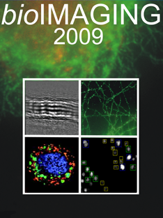
Microscopy has contributed immensely to the development of modern biology since 1665 when Robert Hooke published his book „Micrographia“ depicting a large number of microscopical sketches. In our days a major breakthrough in biology is the discovery of the green fluorescent protein (GFP). Another important innovation was found by the german scientist Stefan Hell from Göttingen. He received for example the „Leibniz Preis“ of the German research community (DFG) for „light microscopy with unknown clarity“. These methods enable the visualization of nanoscopic structures in living cells.
Similar high magnification microscopic plus latest electronmicroscopic techniques are also being developed in the department of physics at the University of Bielefeld. A third building block will lead from August 25-28, 2009 from microscopy to imaging.
The topics include:
– Beyond Optical Microscopy
– High Resolution Microscopy in Biology
– From Life Cell Imaging to Systems Biology
– Bioimaging Informatics
The registration is open until July 11, 2009.
www.cebitec.uni-bielefeld.de/symposium/bioimaging

bioImaging 2009
 Leave a Comment » |
  Events | Verschlagwortet: Bielefeld, Bio, bioimaging, bioImaging 2009, Electronmicroscope, electronmicroscopic, event, GFP, green fluorescent protein, Image Processing, imaging, Methods, Microcoscpy, Microscopica, nanoscopic, optical, University, visualization |
  Permalink
 Veröffentlicht von aszerdi
Januar 9, 2009
From Sunday April 5 to Wednesday April 8, 2009 the Focus on Microscopy (FOM) conference will take place in Krakow, Poland. It is the continuation of a yearly conference series presenting the latest innovations in optical microscopy and its application in biology, medicine and the material sciences. Key subjects are the theory and practice of 3D optical imaging, related 3D image processing, and reporting especially on developments in resolution and imaging modalities. The FOM conference also covers the rapidly advancing fluorescence labeling techniques for the confocal and multiphoton 3D imaging of live- biological-specimens. A technical exhibition will be a special feature of this year’s conference in Krakow.
Upcoming topics will cover:
– Confocal and multiphoton-excitation microscopy
– Novel illumination and detection strategies
– Fluorescence: new labels, fluorescent proteins, quantum dots, single molecule
– Time-resolved fluorescence: FRET, FRAP, FLIM, FCS
– Coherent non-linear microscopy: SHG, THG, SFG, CARS
– Raman, light scattering microscopy
– Multi-dimensional imaging
– Sub-wavelength resolution: near field microscopy, STED, PALM
– Laser manipulation, ablation and microdissection, photoactivation
– Optical tools in genomics, proteomics, phenomics, cytometry
– Magnetic resonance and X-ray microscopy
– Image processing and visualization
– Live cell and whole tissue imaging
The conference will take place at the Jagiellonian University Auditorium Maximum, ul. Krupnicza 35, in the center of Krakow.
Details for registration, abstract submission, deadlines, etc. will soon be available on:
www.focusonmicroscopy.org

Krakow, Poland (source: pixelio.de)
 Leave a Comment » |
  Events | Verschlagwortet: 3D, attendee, conference, confocal, electron, Events, exhibition, FCS, FLIM, fluorescence, focus, FRAP, FRET, genomics, illumination, imaging, Jagiellonian, Krakow, Krupnicza 35, light, magnetic, meeting, microdissection, microscopy, multiphoton, optical, phenomics, photoactivation, processing, proteomics, raman, resolution, resonance, scattering, series, visualization, wavelength |
  Permalink
 Veröffentlicht von aszerdi


 Veröffentlicht von aszerdi
