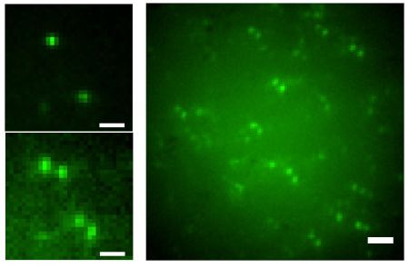
Scientists at Heidelberg University, Germany have developed a new technique for localization microscopy, the “spectral precision distance microscopy” (SPDM). Using visible light, this method allows a single molecule resolution of celullar structures down to the range of few nanometer, about 20 times better than the conventional optical resolution. The researchers invented a new instrument which is a combination of the world’s fastest nano light microscope for 3D cell analysis and the new SPDM technique. Prof. Christoph Cremer of the Kirchhoff Institute of Physics and his team were able to show that SPDM can be realized by common fluorescent dyes, such as the green fluorescent protein (GFP) which can be switched on and off by means of light, as long as certain photophysical conditions are fulfilled. This can be achieved via the so-called “reversible photobleaching” of the dye. So far, only special fluorescent dyes could be used as temporally convertible light signals. According to Cremer there are millions of specimens containing gene constructs with dyes from the GFP group available in biomedical laboratories all over the world. They could be put into immediate use for this new kind of localization microscopy.
www.uni-heidelberg.de
Localization Microscopy Using GFP
Juli 30, 20093D Movies of Microscopic Systems
Juli 29, 2009Physicists at New York University (NYU), US have developed a technique to record three-dimensional movies of microscopic systems, such as biological molecules, through holographic video. The technique, developed in the laboratory of NYU Physics Professor David Grier, is comprised of two components: making and recording the images of microscopic systems and then analyzing these images. To generate and record images, the researchers created a holographic microscope. It is based on a conventional light microscope, which uses a collimated laser beam instead of on an incandescent illuminator.
When an object is placed into path of the microscope’s beam, the object scatters some of the beam’s light into a complex diffraction pattern. The scattered light overlaps with the original beam to create an interference pattern reminiscent of overlapping ripples in a pool of water. The microscope then magnifies the resulting pattern of light and dark and records it with a conventional digital video recorder. Each snapshot in the resulting video stream is a hologram of the original object. Unlike a conventional photograph, each holographic snapshot stores information about the three-dimensional structure and composition of the object that created the scattered light field. The recorded holograms appear as a pattern of concentric light and dark rings.
For analyzing the images the researchers based their work on a quantitative theory, the Lorenz-Mie theory, which maintains that the way light is scattered can reveal the size and composition of the object that is scattering it.
The application of the technique ranges from research in fundamental statistical physics to analyzing the composition of fat droplets in milk.
www.nyu.edu

In the microscope, a laser beam illuminates the sample. Light scattered by the sample creates an interference pattern which is magnified and recorded. Then measurements of the particle’s position, size, and refractive index are obtained.
The Sound of Light
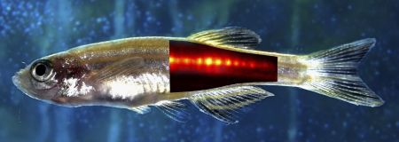
Juli 1, 2009Together with his research team, Professor Vasilis Ntziachristos from the Helmholtz Zentrum Munich, Germany and the Technical University Munich, Germany developed a new technology to make light audible. The technique, called multi-spectral opto-acoustic tomography (MSOT), combines light and ultrasound to visualize fluorescent proteins that are seated several centimeters deep into living tissue.
The researchers used a genetically modified adult zebra fish which carried fluorescent pigments in its tissue. They illuminated the fish from multiple angles using flashes of laser light that are absorbed by the fluorescent pigments in the fish. The pigments absorb the light, a process that causes slight local increases of temperature, which in turn result in tiny local volume expansions. This happens very quickly and creates small shock waves. In effect, the short laser pulse gives rise to an ultrasound wave that the researchers pick up with an ultrasound microphone. To analyze the resulting acoustic patterns, a computer is attached. The computer uses specially developed mathematical formulas to evaluate and interpret the specific distortions caused by scales, muscles, bones and internal organs to generate a three-dimensional image. In the future this technology may facilitate the examination of tumors or coronary vessels in humans.
www.helmholtz-muenchen.de/en

Multi-spectral opto-acoustic tomography or MSOT allows the investigation of subcellular processes in live organisms.
Interdisciplinary Symposium on 3D Microscopy
Juni 3, 20093D Microscopy, taking place from July 12 -16, 2009 in Interlaken, Switzerland, is an international symposium focused on 3D imaging and spectroscopy in science. The objective of this symposium is to create a forum for researchers with different expertise and scientific interests to present their knowhow and the techniques they use to answer their scientific questions. Methods using three-dimensional imaging and spectroscopy to retrieve data in volume will be discussed, whether “light”, x-rays, electrons or near field probes are used. The conference contains 3 plenary talks and 9 sessions with invited and contributed talks and poster sessions.
Session topics are:
– High resolution TEM and AFM
– 3D CLSM and light microscopy
– Stereology
– 3D TEM and atom probe tomography/serial sectioning
– 3D correlative microscopy
– 3D X-ray microscopy and tomography
– 3D FIB/SEM or serial sectioning (with Denk-method) blockface tomography
– 3D image analysis and simulation
– 3D scanning probe microscopy

Interlaken, Switzerland
Frontiers of Electron Microscopy in Materials Sciences
Mai 25, 2009The Twelfth Frontiers of Electron Microscopy in Materials Science, FEMMS2009, will take place from Sept. 27 – Oct. 2, 2009 at “Huis Ten Bosch” in Sasebo/Nagasaki in Kyushu Island, Japan. FEMMS is an international a biennial symposium series focused on the application of electron microscopy, primarily TEM, in the field of materials science. The conference contains a plenary talk, 9 sessions of invited talks and poster sessions of contributed papers. The sessions cover recent progresses and emerging trends, such as current instrument advances in TEM, SEM, HVEM and detecting systems, ultra-high resolution imaging and analysis, in-situ and ultra-fast analysis, 3-dimensional analysis, and so on. Dr. Akira Tonomura, a world renowned pioneer in the field of electron holography, will give a plenary talk as the distinguished lectureship award winner.
www.femms2009.org

3D in 12 Days
April 30, 2009During June, 13-25, the 14th annual Living Cell Course will take place at the University of British Columbia Medicine School (UBC) in Vancouver, Canada. This residential course concentrates on all aspects of the 3D microscopy of living cells. Designed for biological research scientists and advanced graduate students, who apply – or plan to – modern 3D imaging, the course want to open up-to-date methods to a wider selection of scientists. The aim of this intense course is to bring students and manufacturers together. The course’s topics include amongst others scanning systems like AODs, mirrors and disks, deconvolution of wide-field and confocal data, dye design, poisson noise QE and S/N, calcium imaging, as well as „how to keep cells alive“.
www.3dcourse.ubc.ca/index.htm

Vancouver, Canada (sorce: pixelio.de)
Microbeam Analysis – EMAS 2009
April 24, 2009The European Microbeam Analysis Society (EMAS) will hold its 11th annual workshop on „Modern Developments and Applications in Microbeam Analysis“ from May 10-14, 2009 in Gdynia/Rumia, Gdansk, Poland. The main topics are electron probe microanalysis, micro- and nanoanalysis in the natural resources industry, fast energy-dispersive X-ray spectrometry, electron backscatter diffraction, and three-dimensional microanalysis. Time will also be devoted to problem orientated application of microbeam analysis techniques in fields such as catalysts, composites, glass, sensors, and in cultural heritage, environment, forensics, geology, mineralogy, metallurgy, microelectronics, surfaces and interfaces. The event will take place at the Hotel Spa Faltom, Gdynia/Rumia, Gdansk.
www.emas-web.net

City of Gdansk, Poland (source: pixelio.de)
3D Single Molecule Imaging
März 20, 2009
A team of researchers led by professor Rafael Piestun of the department of electrical and computer engineering at the University of Colorado and William E. Moerner, professor of chemistry at Stanford University, have demonstrated for the first time a method for three-dimensional optical imaging of objects smaller than 20 nanometers over a wide spatial range. Optical imaging at these scales is of great interest in biomedical sciences and nanotechnology. The new findings, which provide a powerful tool for the super resolution of single molecules, have implications for characterizing defects in materials, the characterization of nanostructures, and the three-dimensional, biophysical and biomedical imaging of tagged molecules inside and outside of cells.
www.stanford.edu
www.colorado.edu

3D single molecule imaging
The Smallest Periscope
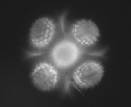
März 11, 2009A team of Vanderbilt scientists have invented the world’s smallest version of the periscope and are using it to look at cells and other microorganisms from several sides at once. The researchers have dubbed their devices „mirrored pyramidal wells.“ They consist of pyramidal-shaped cavities molded into silicon whose interior surfaces are coated with a reflective layer of gold or platinum. They are in dimension about the width of a human hair and can be made in a range of sizes to view different-sized objects. When a cell is placed in such a well and viewed with a regular optical microscope, the researcher can see not only the top of the cell, but several sides simultaneously. „This is something biologists almost never see,“ says team member Chris Janetopoulos, assistant professor of biological sciences.
The Vanderbilt group is not the first to make microscopic pyramidal wells, but it is the first to apply them to make 3D images of microorganisms. In 2006, a group of scientists in England created pyramidal micromirrors and applied them to trapping atoms. And last spring researchers at the National Institute of Standards and Technology used similar structures to track nanoparticles.
www.vanderbilt.edu

Sunflower pollen, taken by Kevin Seale, Vanderbilt Institute for Integrative Biosystems Research and Education
FEI Expands Presence in Mineralogy Market
Januar 15, 2009FEI Company has acquired substantially all of the assets of Intellection Holdings of Brisbane, Australia. Intellection’s primary product is the Qemscan automated mineralogy system. The purchase increases FEI’s presence in the automated mineralogy market for mining companies. The purchase price was approximately $2.8 million. Automated mineralogy systems identify minerals in polished sections of drill core, particulate, or lump materials and quantify a wide range of characteristics, such as mineral abundance, grain size, and liberation.
www.fei.com
www.intellection.com.au


 Veröffentlicht von aszerdi